
İçöz, Kutay
Loading...
Profile URL
Name Variants
Icoz, Kutay İçöz, Kutay
Job Title
Email Address
Main Affiliation
01. Abdullah Gül University
Mühendislik Fakültesi
Elektrik-Elektronik Mühendisliği
Mühendislik Fakültesi
Elektrik-Elektronik Mühendisliği
Status
Former Staff
Website
ORCID ID
Scopus Author ID
Turkish CoHE Profile ID
Google Scholar ID
WoS Researcher ID
Sustainable Development Goals

This researcher does not have a Scopus ID.

This researcher does not have a WoS ID.
No records found in other affiliations.
| Journal | Count |
|---|
Current Page: 1 / NaN
Scopus Quartile Distribution
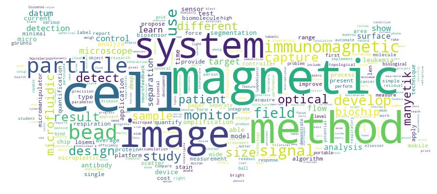
Competency Cloud
46 results
Scholarly Output Search Results
Now showing 1 - 10 of 46
Conference Object Image-Processing Based Signal Readout Method For MRD Biochip(Institute of Electrical and Electronics Engineers Inc., 2019-04) Uslu, Fatma; İçöz, Kutay; Taşdemir, KasímThe response of the cancer patients to chemotherapy treatment varies from person to person. For some patients cancer cells are resistant to treatment and these cells can relapse again which is known as minimal residual disease. A microfluidic-based biochip capable of monitoring minimal residual disease is under development by our research group. The role of the biochip is to capture the target cells, which were separated by immunomagnetic beads on micro square tiles. Then biochips are imaged using a bright field optical microscope and it is planned to perform image-processing methods to detect the target cells, immunomagnetic beads and micro tiles. In this work the current progress of image processing methods for differentiating the immunomagnetic beads and micro tiles is presented. © 2020 Elsevier B.V., All rights reserved.Article Citation - WoS: 3Citation - Scopus: 6Simple Staining of Cells on a Chip(MDPI, 2022-11-13) Kosker, Fatma Betul; Aydin, Omer; Icoz, KutaySimple staining of cells is a widely used method in basic medical diagnostics, education, and research laboratories. The stains are low-cost, but the extensive consumption results in excessive toxic waste generation. Thus, to decrease the amount of toxic waste resulting from the cell staining procedure is a need. In this study, we developed a magnetically driven and compartmentalized passive microfluidic chip to perform simple staining of human eukaryotic cells, K562 cells, and lymphocyte cells derived from patients. We demonstrated simple staining on cells with trypan blue, methylene blue, crystal violet, and safranin for high, medium, and low cell densities. The stained cells were imaged using a bright field optical microscope and a cell phone to count cells on the focal plane. The staining improved the color signal of the cell by 25-135-pixel intensity changes for the microscopic images. The validity of the protocol was determined using Jurkat and MDA-MB-231 cell lines as negative controls. In order to demonstrate the practicality of the system, lymphocyte cells derived from human blood samples were stained with trypan blue. The color intensity changes in the first and last compartments were analyzed to evaluate the performance of the chip. The developed method is ultra-low cost, significantly reduces the waste generated, and can be integrated with mobile imaging devices in terms of portability. By combining microfabrication technology with cell staining, this study reported a novel contribution to the field of microfluidic biosensors. In the future, we expect to demonstrate the detection of pathogens using this method.Article Citation - WoS: 14Citation - Scopus: 16Detection of Proteins Using Nano Magnetic Particle Accumulation-Based Signal Amplification(MDPI, 2016-11-29) Icoz, Kutay; Mzava, OmaryWe report a biosensing method based on magnetic particles where coated magnetic particles are used for immunomagnetic separation, and uncoated magnetic particles are used for signal enhancement. To quantify the signal amplification, optical micrographs are analyzed to measure changes in pixel area and pixel intensity. Microcontact-printed surface receptors are arranged in alternating lines on gold chips, enabling differential calculations. In a model experiment, target molecules-streptavidin-are first captured and separated by biotin-coated magnetic particles, and then exposed to a gold surface functionalized with biotin-coupled bovine serum albumin, forming a sandwich assay. Applying a magnetic field and introducing uncoated magnetic particles resulted in accumulation around magnetic particles in the sandwich assay and enhancement of the contrast to noise ratio at least by eight-fold in a range of 0.1-100 mu M.Conference Object A Horizontal Magnetic Tweezer for Single Molecule Micromanipulations(Institute of Electrical and Electronics Engineers Inc., 2018-10) Ablay, Günyaz; Böyük, Mustafa; Eroǧlu, Yakup; İçöz, KutayMagnetic tweezers are able to manipulate cells or biomolecules for various applications and measurements. In this work, a horizontal magnetic tweezer is designed, modeled and controlled for single molecule manipulations. A method is presented for dynamic modeling of horizontal magnetic tweezers. A linear control method is designed to ensure a wide range of operation conditions with zero steady-state error. The horizontal magnetic tweezer is able to generate a wide range of piconewton scale forces on a superparamagnetic microparticle for single molecule separation, and biosensor developments. © 2019 Elsevier B.V., All rights reserved.Master Thesis Hedeflenen Lösemik Hücrelerin Sayımı için Görüntü İşleme Tabanlı Ölçüm Sistemi(Abdullah Gül Üniversitesi, 2019) TAŞ, ZEHRA; Taş, Zehra; İçöz, KutayAkut Lenfosit Lösemi (ALL) çocukluk çağında en sık görülen kanser türüdür. Bu kanserin ilerlemesi oldukça hızlıdır, bu yüzden tedavi için zaman çok önemlidir. Hastalar için ilaç tedavisi (kemoterapi), kemik iliği nakli, radyasyon tedavisi ve immünoterapi gibi bazı tedaviler vardır. Bu tedaviler arasında kemoterapi öncelikle tercih edilen bir yöntemdir, ancak sonucu hastadan hastaya farklılık göstermektedir. Hastaların iyileşmesinde ilacın dozajını ayarlamak için kemoterapinin tedaviye etkisini ölçmek çok önemlidir. Kemoterapinin etkisini ölçmek için akım sitometrisi (FC), polimeraz zincir reaksiyonu (PCR) ve mikroskobik inceleme teknikleri kullanılır. Ancak, bu yöntemler zaman alır, pahalıdır ve uzman gerektirir. Bu tezde, sorun görüntü işleme tekniklerine dayanan otomatik bir hücre algılama ve niceleme yöntemi ile ele alınmaktadır. Optik mikroskopi ile elde edilen görüntüler işlenir ve immüno-manyetik boncuklarla yakalanan ALL hücrelerinin ölçümü sağlanır. Geleneksel yöntemlerle karşılaştırıldığında, bu teknik zaman verimli, ucuz ve kullanımı kolaydır. ALL hücrelerinin miktarının yanı sıra, biyosensörlerin sinyal büyütme metodu için projenin ilk bölümünde kullanılan manyetik boncukların belirlenmesi için görüntü işleme algoritmaları da geliştirilmiştir. Görüntüler cep telefonu mikroskopisinden hem mikroskop lamı hem de difraksiyon ızgarası tabanlı biyosensörler için elde edilmiştir. Her iki yöntemde de manyetik boncuk birikimleri gözlenmiş ve geliştirilen algoritmalar cep telefonu mikroskopu görüntülerine uygulanmıştır. Böylece, manyetik boncukların birikimleri bilgisi, görüntü işleme kullanılarak otomatik olarak elde edilmiştir.Article Citation - WoS: 5Citation - Scopus: 6Design, Modeling, and Control of a Horizontal Magnetic Micromanipulator(Sage Publications Ltd, 2019-01-31) Ablay, Gunyaz; Boyuk, Mustafa; Icoz, KutayMagnetic micromanipulators with a wide range of force generating capabilities are able to manipulate micron size particles for various applications and measurements. These magnetic particles can be coated with receptors to specifically bind to target biomolecules. In this work, a horizontal magnetic micromanipulator is designed, modeled and controlled for single micron size magnetic particle manipulations. A method is presented for dynamic modeling of magnetic micromanipulators. A feedback control method is designed that allows direct linearization of the system. It is shown that the proposed controller guarantees the stability of the closed-loop system, and yields zero steady-state error in a wide range of operation conditions. We show that the micromanipulator is able to generate a wide range of piconewton (pN) scale forces on a superparamagnetic particle for single molecule separation, and biosensor developments.Article Citation - WoS: 15Citation - Scopus: 17Numerical Analysis and Experimental Verification of Optical Scattering From Microplastics(Royal Soc, 2023-08) Genc, Sinan; Icoz, Kutay; Erdem, TalhaAccurate and fast characterization of the micron-sized plastic particles in aqueous media requires an in-depth understanding of light interaction with these particles. Due to the complexity of Mie scattering theory, the features of the scattered light have rarely been related to the physical properties of these tiny objects. To address this problem, we reveal the relation of the wavelength-dependent optical scattering patterns with the size and refractive index of the particles by numerically studying the angular scattering features. We subsequently present a low-cost setup to measure the optical scattering of the particles. Theoretical investigation shows that the angular distribution of the scattered light by microplastics carries distinct signatures of the particle size and the refractive index. The results can be used to develop a portable, low-cost setup to detect microplastics in water.Master Thesis Micro Manyetik ve Parçacıklarının Manipülasyonuna Yönelik Sistem Tasarımı(Abdullah Gül Üniversitesi, 2018) BÖYÜK, MUSTAFA; Böyük, Mustafa; İçöz, Kutay; Ablay, GünyazManyetik cımbızlar çeşitli uygulamalar ve ölçümler için hücreleri veya biyomolekülleri manipüle edebilir. Tek moleküllü manipülasyonlar için bir elektromanyetik mikro manipülatör tasarlandı, modellendi ve kontrol edildi. Elektromanyetik cımbız mikron boyutlu süperparamanyetik parçacıkları uygun kontrol mekanizması yardımıyla kontrol edebilir. Bu parçacıklar, hedef biyomoleküllerin yakalanması için reseptörler ile fonksiyonel hale getirilebildiğinden dolayı yüklü partiküller harici bir manyetik alan kullanılarak belirli bir yere taşınabilirler. Manyetik tek kutup ve manyetik devre yaklaşımları, manyetik sistemin dinamik denklemini modellemek için bu çalışmada kullanıldı. Ofset akım tabanlı geri besleme ile doğrusallaştırma yaklaşımı bir kontrolör tasarlanarak, sıfır kararlı-durum hatasıyla geniş çalışma koşulları sağlandı. Tek parçacığın konumunu bulmak için görüntü tabanlı algoritma geliştirilmiştir. Türetilmiş model ve kontrol sistemini doğrulamak için sayısal simülasyonlar yapılır. Tasarlanan manyetik sistem, 1 ila 10 mikrometre çapındaki manyetik bir parçacığı 1 amperden az bir akımla kontrol etmek için 1-100 pN arasında kuvvet uygulayabilmektedir. Manyetik yönlendirici sistemi, tek hücre ayrımı, ve biyosensör gelişmeleri için kullanılabilir.Conference Object Magnetic-Particle Based Signal Amplification Method Integrated With Mobile-Devices for Low Cost Biosensing(Elsevier Science Bv, 2017) Mzava, Omary; Tas, Zehra; Lafci, Vahit Can; Cakar, Mehmet Akif; Ozdur, Ibrahim; Icoz, KutayWe present a signal amplification method for biosensing applications using magnetic particles. In this method, mobile devices and simple spherical glass beads are used as a low-cost microscope to detect magnetic particles. Magnetic particles have two main functions; 1) conventionally capture, separate and transport target molecules 2) form magnetic dipoles under an applied external magnetic field to attract other magnetized particles. When magnetic particles accumulate and form a cluster, the corresponding pixel area in the image taken by the simple microscope is increased resulting in signal amplification. Current focus of new generation biosensor research is to increase the sensitivity levels of the devices to compete with current lab analysis tools while inherently having other advantages such as being low-cost, portable and simple. Biosensors based on micro/nano magnetic particles use various measurement techniques and amplification methods. In order to fully benefit from the advantages of micro/nano technology based systems, measurement set up must be also portable and have high sensitivity. Mobile devices and applications are taking place in medical fields and have high potential for future. In this work mobile devices are employed as measurement setups for the magnetic particle based sensing and signal amplification. The amplification method is not based on bimolecular binding thus cost efficient. After the images of the magnetic particles are taken, these images are sent to cloud computing for analysis by the mobile device. Matlab codes run on cloud servers for processing the images. Finally results are received and displayed on the mobile device. The mobile device based imaging system is able to detect 7 mu m size particles within a 1500 mu m x1500 mu m area and magnetic bead accumulation resulted in at least 5-fold signal amplification. The applied magnetic field is approximately 15 mT and the cost of the system excluding mobile device is under 20 cents. The method is promising for immunomagnetic bead assisted biosensors. (c) 2016 The Authors. Published by Elsevier Ltd.Article Citation - WoS: 3Citation - Scopus: 5Feedback Controller Designs for an Electromagnetic Micromanipulator(Sage Publications Ltd, 2019-09-09) Boyuk, Mustafa; Eroglu, Yakup; Ablay, Gunyaz; Icoz, KutayMagnetic micromanipulators are capable of generating wide range of magnetic forces to manipulate magnetic microparticles for biomedical applications. In this study, a multipole magnetic micromanipulator system including electromagnets, driver circuitry and control unit is designed, modeled and implemented. The micromanipulator can produce a broad range of magnetic forces up to 25 pN on a single magnetic microparticle (1-10 mu m diameter) that is 5 mm away from the electromagnet core tip. Both linear and nonlinear controllers are designed and implemented, and the proposed nonlinear controller produces smooth control currents to assure closed-loop stability of the system with 1 s non-overshoot transient response and zero steady-state tracking error. The maximum output current of the driver circuitry is set to 1 A. The single particle at the center is moved at a speed of 5 mm/s. The fully automatic system can be utilized in applications related to single cell or microparticle manipulations.